Overview
What is a myocardial infarction?
A myocardial infarction is more commonly known as a heart attack, which is permanent damage to heart muscle. It is caused by a loss of blood supply to part of the heart by a blockage in one of the coronary arteries. This leads to damage or death of heart muscle cells and in severe cases, death of the patient.
Brief anatomy of the heart
In our chest the heart sits slightly off to the left and is made purely of muscle which contracts to pump blood all around our bodies. The heart has 3 layers: going from the outermost in, the visceral layer, the myocardium and the endocardium. It is made up of four chambers: 2 superior atria and 2 inferior ventricles. Blood enters the atria and leaves through the ventricles.
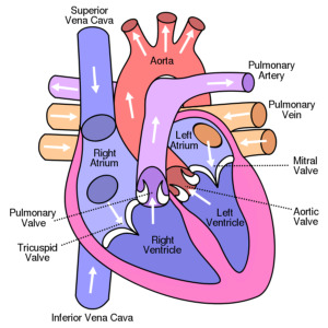 In mammals, there are two circulatory systems, one supplying the lungs (Pulmonary circulation) and one supplying the body (Systemic circulation). Oxygenated blood from the lungs enters the left atrium via the pulmonary vein, the blood then moves into the left ventricle and is pumped around the body via the aorta. Deoxygenated blood returns to the heart via the superior and inferior vena cavae and enters the right atrium. After flowing into the right ventricle, the blood travels to the lungs to receive oxygen via the pulmonary artery.
On both sides of the heart, the ventricles are larger and stronger than the atria as they need more power in order to pump blood out of the heart. The left ventricle is larger than the right due to its role of pumping blood all around the body.
The heart is constantly contracting so needs a constant supply of blood and oxygen. It is the coronary arteries that supply this oxygen and nutrient rich blood to the heart. There are two coronary arteries, left and right, which emerge from the beginning of the aorta near the top of the heart and then branch to penetrate the heart supplying it with oxygenated blood. This circulation is the only source of blood supply to the heart so blockage of it is critical.
Anatomy of the heart image is courtesy of Wikimedia Commons and is thus free of any copyright restrictions.
What happens during a myocardial infarction?
 During a heart attack, a coronary artery or one of its branches is suddenly blocked (see causes). The part of the heart that this artery supplies will lose its blood, oxygen and nutrient supply (ischemia) and unless this blockage is quickly removed, this heart muscle will die affecting the overall function of the heart.
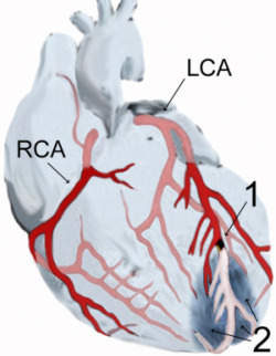
The image on the right is showing a myocardial infarction. (1) shows the location of the occlusion on a branch of the left coronary artery (LCA) (RCA= right coronary artery). (2) shows the myocardial infarction at the tip of the anterior wall of the heart.
The amount of damage done to the heart muscle depends on the size of the area supplied by the blocked artery. If a main coronary artery is occluded, a large area of heart muscle will be in danger whereas if a small branch is blocked, less heart tissue will be affected.
If the disrupted blood flow is not treated immediately, heart muscle cells in the occluded area will die and will be replaced by a collagen scar. If the patient survives, their heart will be permanently damaged. Injured heart tissue conducts electrical impulses more slowly than normal heart tissue meaning that the contracting and pumping ability of the heart will be lessened which can lead to life threatening arrhythmias and possibly the formation of a ventricular aneurysm.
If an MI is not treated, it can lead to heart failure and cardiac arrest in which the patient will not survive.
Myocardial Infarction image is courtesy of Wikimedia Commons and is thus free of any copyright restrictions.
Classification of MIs
There are two types of myocardial infarction based on the location of the coronary artery blockage:
- Transmural: Blockage of a major coronary artery. These infarcts extend through the whole thickness of the heart muscle due to complete blockage of the area’s blood supply.
- Subendocardial: This infarction affects only heart muscle below the endocardium and is due to a local decrease in blood supply.
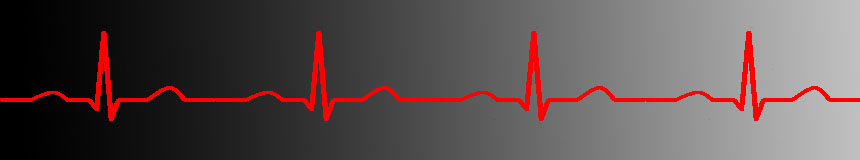
Clinically, MI is further subdivided into ST elevation and non-ST elevation based on ECG changes (see Diagnosis and Treatment for ECG explanation).
A non-ST elevated MI does not cause any changes to the ECG; instead it is detected by chemical changes in the blood. It is usually caused by a partial or temporary blockage in the coronary artery.
An ST elevated MI does cause a change to the ECG reading as well as chemical changes in the blood. This type of MI is more severe as it is caused by a prolonged blockage in the coronary arteries and affects a larger area of the heart.